Projects
Three-Dimensional Visualization
Automated analysis and visualization of three-dimensional, intact-tissue transcriptomic image data
PI: Viviana Gradinaru (Division of Biology and Biological Engineering)
SASE: Julia Vendemiatti, Scholar
Spatial transcriptomic analysis of biological tissue samples aims at attaining the gene expression profiles of numerous cells by detecting hundreds to thousands of RNA transcripts in situ. This approach has provided a unique means to comprehensively understand the genetic identities or functional states of the cells with preservation of their spatial organization in tissue. While most relevant studies have focused on targeting endogenous genes, the Gradinaru lab has been expanding this approach to enable detection of virally delivered transgenes and screening a pool of adeno-associated viruses (AAVs) along with endogenous cell markers in intact tissue, aiming at developing AAV-based, targeted gene delivery systems.
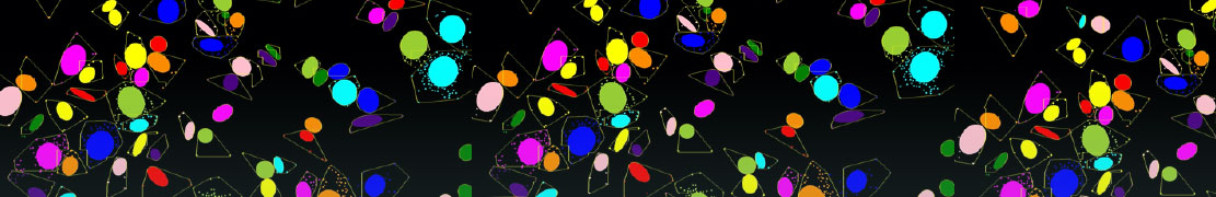
Intact-tissue transcriptomic experiments either for endogenous or exogenous genes rely on three-dimensional, high-resolution confocal imaging that results in massive image datasets. These data usually include RNA signals as diffraction-limit spots (hundreds of nanometers in diameter), nuclei, and sometimes cell boundaries to aid cell segmentation. Although many image processing toolboxes are currently available, there has been a lack of computational solutions designed for such data that would require accurate segmentation of individual cells and their RNA spots in 3D.
To address this, the Lab developed their our own python-based image processing pipeline for cell segmentation and RNA spot detection. The Schmidt Academy collaborated with the Lab on an upgrade the data analysis pipeline by making it much more modular, documented, and user-friendly. This pipeline will be made public for other labs to use.
