Projects
Computational Microscopy: A new way to integrate hardware and algorithms
Haowen Zhou (Division of Engineering and Applied Sciences), SASE Graduate Fellow
The field of optical microscopy is essential for biomedical research and clinical diagnostics, providing critical insights into cellular and tissue structures. However, the advancement of traditional optical hardware is increasingly limited by physical constraints. Computational microscopy, which integrates algorithms with optical systems, offers a solution by overcoming these limitations and enhancing the performance of existing microscopes. The Caltech Biophotonics Lab, led by Prof. Changhuei Yang, invented Fourier ptychographic microscopy (FPM), a next-generation technique that employs complex-field imaging, high spatial-bandwidth, and digital aberration correction. These innovations enable high-quality, high-resolution imaging, which are particularly valuable in fields like digital pathology, label-free cell imaging and microbiology.
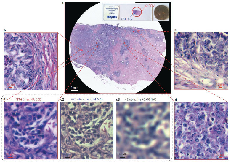
Figure caption: Fourier ptychographic microscopy achieves large field-of-view and high-resolution simultaneously. Image Courtesy of Zheng et al. Nat. Photonics (2013).
To bridge the gap between complex image reconstruction algorithms and practical usage in clinical settings, in the collaboration with Schmidt Academy, the group aims to develop a software platform that unifies existing computational microscopy algorithms. The software will integrate diverse algorithms into a single system that is adaptable to various microscopy setups. This will allow pathologists and medical trainees to seamlessly process large batches of high-resolution histological images without needing to understand the underlying complex computational methods.
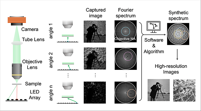
Figure caption: Principle of Fourier Ptychographic Microscopy.
One of the primary challenges of this project is the integration of various image reconstruction algorithms into a flexible, user-friendly platform that works with different optical hardware configurations. This requires creating an adaptable software architecture that can automatically select the best algorithm based on the sample type, microscope setup, and user requirements. Ensuring this platform is intuitive for non-expert users, such as pathologists, adds another layer of complexity. By adhering to best practices in software engineering—such as modular design, robust documentation, and thorough testing—the software will offer a reliable and user-friendly interface. The ultimate goal is to facilitate the adoption of computational microscopy in both research and clinical practice, empowering medical professionals to make faster and more accurate diagnoses. This software platform will not only advance the state of the art in image reconstruction algorithms but also help to accelerate the commercialization of computational microscopy systems.
The Schmidt Academy collaborated with the Caltech Biophotonics Lab to develop a user-friendly software platform that bridges advanced computational microscopy algorithms with practical clinical and research workflows. While a collection of standalone scripts for FPM and related reconstruction methods already existed, they lacked the structure, usability, and robustness required for widespread adoption by non-expert users such as pathologists and medical trainees. To address this, a unified and modular software platform was developed to integrate diverse computational microscopy algorithms into a single, adaptable system compatible with multiple microscope configurations. The platform emphasizes best practices in software engineering, including modular design, configurability, thorough documentation, and testing, enabling seamless processing of large-scale, high-resolution imaging datasets without requiring users to understand the underlying computational complexity. Beyond supporting FPM, this software framework is designed to be extensible to other computational microscopy techniques, accelerating both research translation and clinical deployment. Through this collaboration, the lab has also strengthened its software development culture by centralizing codebases on GitHub and adopting modern software engineering practices, thereby facilitating long-term maintenance, collaboration, and commercialization of computational microscopy technologies.
GitHub repository: https://github.com/hwzhou2020/FPM_software
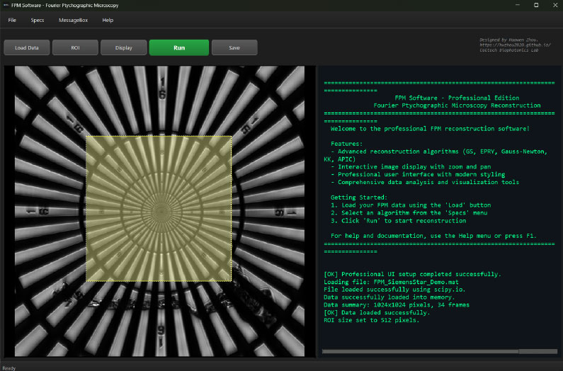
Figure caption: A screenshot of the software interface.
Cover picture: